Olivia has a clinical diagnosis of Goldblatt Syndrome. The main symptoms are short stature, joint laxity, muscle weakness, narrow chest, and dentinogenesis imperfecta (weak tooth enamel).
Mommy takes me to appointments with doctors all over the world to help me feel better. I have Physiotherapy, Occupational Therapy, Hydrotherapy several times a week. I’m an extremely clever girl, but I suffer everyday from pain and this makes me sad. Thank you for following my journey.
Below is the medical information about my syndrome (this information was found on the OMIM website: https://www.omim.org/entry/184260)
Description of Spondylometaphyseal Dysplasia with Dentinogenesis Imperfecta/ Odontochondrodysplasia (ODCD)
Alternative titles: Goldblatt Syndrome
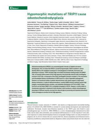
The 2019 published article titled “Hypomorphic mutations of TRIP11 cause odontochondrodysplasia” will offer detailed information on the genetic Origin of Goldblatt Syndrome. Click HERE to read this article.
Clinical Features
Key clinical findings are short stature, narrow chest, mesomelic limb shortening, brachydactyly, joint laxity, and dental anomalies. Radiographic features include platyspondyly with coronal clefts and metaphyseal irregularities of the tubular bones.
In a 3.5-year-old boy of mixed Australian/Thai parentage, Goldblatt et al. (1991) described a form of spondylometaphyseal dysplasia associated with joint laxity and dentinogenesis imperfecta. The disorder appeared to be different from the spondyloepimetaphyseal dysplasia with joint laxity described by Beighton et al. (1984) (see 271640) as well as from the forms of spondylometaphyseal dysplasia known as the Irapa (271650) and Strudwick (184250) types. The dentinogenesis imperfecta resembled that seen with osteogenesis imperfecta but there were no signs of that disorder. The father’s age was not given. X-linked spondylometaphyseal dysplasia (313420) is not associated with dentinogenesis imperfecta.
Bonaventure et al. (1992) presented the cases of 2 additional patients: 2 sibs with short limb dwarfism, joint laxity, and dentinogenesis imperfecta, born to healthy nonconsanguineous parents. Electron microscopic studies revealed large vacuoles of dilated rough endoplasmic reticulum within the cytoplasm of chondrocytes. Gel electrophoresis of pepsin-soluble collagen extracted from cartilage demonstrated type II collagen chains with abnormal mobility. A quantitative decrease in COL1A1 and COL1A2 mRNA was also observed. Overmodification of type II collagen peptides, suggested by the findings, was consistent with the presence of a single base substitution in the COL2A1 gene (120140).
Unger et al. (2008) reported 6 additional patients with Goldblatt syndrome, including a second sib pair (brother and sister). The main clinical signs included short stature, joint laxity, narrow chest, and dentinogenesis imperfecta. Two patients required spinal surgery to correct progressive scoliosis at age 4.5 years and 7 years. Intellectual development was normal but motor delays occurred in some patients. The main radiographic features included congenital platyspondyly with coronal clefts, severe metaphyseal changes (especially hands, wrists, and knees), mesomelic limb shortening, and coxa valga. The occurrence of a second sib pair suggests autosomal recessive inheritance, although none of the reported families (including those with sib recurrence) were consanguineous. One patient was initially suspected of having Torrance type platyspondylic lethal skeletal dysplasia (PLSDT; 151210).
Molecular Genetics
Unger et al. (2008) performed complete sequencing of several genes, including the COL2A1 gene, in one or more the patients they reported with Goldblatt syndrome and identified no pathogenic mutations.
2022 Xrays of Olivia Lugani


Medical information
Below some articles which explains in more details my syndrome and what it does to the body.
The article below explains the genetic origin of the disorder (click HERE to read article).
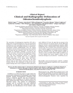
The article below explains Goldblatt syndrome (click HERE or on the image).
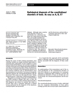
The article below explains the bone disorder (click HERE or on the image).